lv summit pvc morphology | cardiac ablation pvc news today lv summit pvc morphology In general, there is a gradual progression in V 1 morphology from a complete LBBB to an RBBB as the SOO shifts from anterior to posteriorly directed structures (ie, anterior . Africa Inland Mission (AIM) is a Christian mission sending agency with a heart for Africa’s peoples. We have our roots in a small band of faithful men and women who, in 1895, .
0 · ventricular ectopics ecg images
1 · summit pvc location
2 · rvot free wall pvc
3 · right ventricular outflow tract pvcs
4 · right ventricular outflow tract anatomy
5 · lv summit pvc ablation
6 · cardiac ablation pvc news today
7 · aortomitral continuity pvc
$100.00
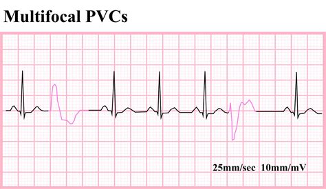
ventricular ectopics ecg images
la femme prada fragrantica
In general, there is a gradual progression in V 1 morphology from a complete LBBB to an RBBB as the SOO shifts from anterior to posteriorly directed structures (ie, anterior .Background: Several algorithms have been proposed to predict the origin of outflow .Background— The summit of the left ventricle (LV) is the most superior . The initial 12-lead electrocardiogram showed a PVC with left bundle branch block morphology and inferior axis with an early transition in lead V 3 (Figure 1 a). The PVC .
The second PVC is a clinical PVC (marked by the arrow) with an RBBB pattern, a right inferior axis, and a QS pattern in lead I. The first PVC is a catheter-induced PVC, while .
Background— The summit of the left ventricle (LV) is the most superior portion of the epicardial LV bounded by an arc from the left anterior .Background—The summit of the left ventricle (LV) is the most superior portion of the epicardial LV bounded by an arc from the left anterior descending coronary artery, superior to the first septal . We describe the prevalence, characteristics, and outcomes of a new distinct ECG pattern of outflow tract ventricular arrhythmias (OTVAs) with left bundle branch morphology and abrupt transition in lead V 3 (ATV3) that .This region is the highest portion of the LV epicardium, near the bifurcation of the left main coronary artery (LMCA), and accounts for up to 14.5% of LV VAs.2 The complex relationships .
The initial 12-lead electrocardiogram showed a PVC with left bundle branch block morphology and inferior axis with an early transition in lead V3 (Figure 1a). The PVC displayed a QRS duration .
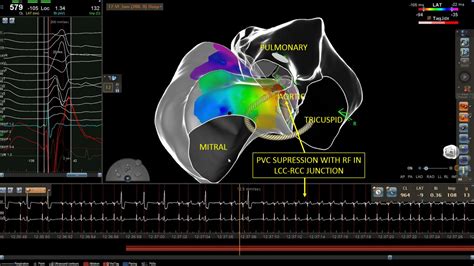
We describe a LV Summit PVC originated from its medial and superior epicardial aspect. Description of wire mapping technique and ablation approach is provided.The left ventricular (LV) summit can be the location of idiopathic ventricular arrhythmias. The LV summit is the most superior portion of the epicardial LV outflow tract area bounded by the left . In general, there is a gradual progression in V 1 morphology from a complete LBBB to an RBBB as the SOO shifts from anterior to posteriorly directed structures (ie, anterior RVOT to posterior RVOT, RCAS-LCAS commissure, LCAS, AMC, great cardiac vein/anterior interventricular vein, and LV summit).
summit pvc location
The initial 12-lead electrocardiogram showed a PVC with left bundle branch block morphology and inferior axis with an early transition in lead V 3 (Figure 1 a). The PVC displayed a QRS duration of 148 ms, a QS pattern in lead I, a maximum deflection index of 0.8, an intrinsicoid deflection time of 52 ms, and an aVL/aVR Q-wave ratio of 1.6. The second PVC is a clinical PVC (marked by the arrow) with an RBBB pattern, a right inferior axis, and a QS pattern in lead I. The first PVC is a catheter-induced PVC, while the ablation catheter is located in the LV subvalvular portion of the LCC, which has a different morphology from the clinical PVC. The pace map from this site is also poor. Background— The summit of the left ventricle (LV) is the most superior portion of the epicardial LV bounded by an arc from the left anterior descending coronary artery, superior to the first septal perforating branch to the left circumflex coronary artery.Background—The summit of the left ventricle (LV) is the most superior portion of the epicardial LV bounded by an arc from the left anterior descending coronary artery, superior to the first septal perforating branch to the left circumflex coronary artery.
We describe the prevalence, characteristics, and outcomes of a new distinct ECG pattern of outflow tract ventricular arrhythmias (OTVAs) with left bundle branch morphology and abrupt transition in lead V 3 (ATV3) that localizes to the septal margin of the LV summit.
This region is the highest portion of the LV epicardium, near the bifurcation of the left main coronary artery (LMCA), and accounts for up to 14.5% of LV VAs.2 The complex relationships between the left ventricular summit (LVS) and surrounding structures under-score the importance of understanding the anatomy of this region and the value of imag.The initial 12-lead electrocardiogram showed a PVC with left bundle branch block morphology and inferior axis with an early transition in lead V3 (Figure 1a). The PVC displayed a QRS duration of 148 ms, a QS pattern in lead I, a maximum de ection index of 0.8, fl. an intrinsicoid de ection time of 52 ms, and an aVL/aVR. fl. Q-wave ratio of 1.6.
We describe a LV Summit PVC originated from its medial and superior epicardial aspect. Description of wire mapping technique and ablation approach is provided.The left ventricular (LV) summit can be the location of idiopathic ventricular arrhythmias. The LV summit is the most superior portion of the epicardial LV outflow tract area bounded by the left anterior descending (LAD) and left circumflex (LCx) arteries and the great cardiac vein (GCV). In general, there is a gradual progression in V 1 morphology from a complete LBBB to an RBBB as the SOO shifts from anterior to posteriorly directed structures (ie, anterior RVOT to posterior RVOT, RCAS-LCAS commissure, LCAS, AMC, great cardiac vein/anterior interventricular vein, and LV summit). The initial 12-lead electrocardiogram showed a PVC with left bundle branch block morphology and inferior axis with an early transition in lead V 3 (Figure 1 a). The PVC displayed a QRS duration of 148 ms, a QS pattern in lead I, a maximum deflection index of 0.8, an intrinsicoid deflection time of 52 ms, and an aVL/aVR Q-wave ratio of 1.6.
The second PVC is a clinical PVC (marked by the arrow) with an RBBB pattern, a right inferior axis, and a QS pattern in lead I. The first PVC is a catheter-induced PVC, while the ablation catheter is located in the LV subvalvular portion of the LCC, which has a different morphology from the clinical PVC. The pace map from this site is also poor. Background— The summit of the left ventricle (LV) is the most superior portion of the epicardial LV bounded by an arc from the left anterior descending coronary artery, superior to the first septal perforating branch to the left circumflex coronary artery.
Background—The summit of the left ventricle (LV) is the most superior portion of the epicardial LV bounded by an arc from the left anterior descending coronary artery, superior to the first septal perforating branch to the left circumflex coronary artery. We describe the prevalence, characteristics, and outcomes of a new distinct ECG pattern of outflow tract ventricular arrhythmias (OTVAs) with left bundle branch morphology and abrupt transition in lead V 3 (ATV3) that localizes to the septal margin of the LV summit.This region is the highest portion of the LV epicardium, near the bifurcation of the left main coronary artery (LMCA), and accounts for up to 14.5% of LV VAs.2 The complex relationships between the left ventricular summit (LVS) and surrounding structures under-score the importance of understanding the anatomy of this region and the value of imag.The initial 12-lead electrocardiogram showed a PVC with left bundle branch block morphology and inferior axis with an early transition in lead V3 (Figure 1a). The PVC displayed a QRS duration of 148 ms, a QS pattern in lead I, a maximum de ection index of 0.8, fl. an intrinsicoid de ection time of 52 ms, and an aVL/aVR. fl. Q-wave ratio of 1.6.
We describe a LV Summit PVC originated from its medial and superior epicardial aspect. Description of wire mapping technique and ablation approach is provided.


Manuals and User Guides for AHT MALTA Series. We have 2 AHT MALTA Series manuals available for free PDF download: Operating Manual, Instruction Manual. AHT MALTA Series Operating Manual (27 pages) Commercial refrigerating appliance. Brand: AHT | Category: Refrigerator | Size: 1.09 MB. Table of Contents. Model Types - .
lv summit pvc morphology|cardiac ablation pvc news today


